Imagine entering a clinic where a damaged valve, patch of skin, or section of cartilage is not harvested from another person but assembled inside a machine, layer by layer, from the patient’s own biological material. That scene no longer belongs to science fiction. 3D bioprinting is moving from laboratory demonstrations toward clinical use, offering a new way to repair tissue, test therapies, and perhaps address one of medicine’s most stubborn problems: the shortage of compatible donor organs.
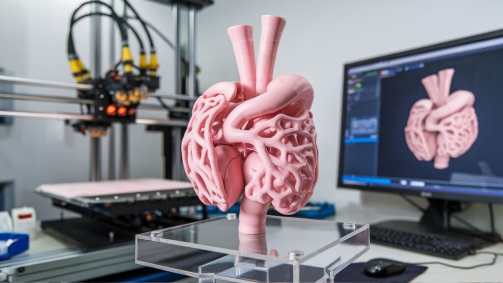
At its core, bioprinting is a manufacturing process that uses living cells and supportive biological materials instead of plastic or metal. These materials, often called bio-inks, are deposited in precise patterns to form three-dimensional structures that can behave like real tissue. The goal is not merely to create a shape, but to reproduce the cellular architecture, mechanical properties, and biological function needed for healing.
The approach differs from conventional tissue engineering in important ways. Traditional methods may rely on scaffolds, tissue expansion, or grafts taken from other sites in the body. Bioprinting can place cells in defined positions, which may improve vascularization, structural accuracy, and patient-specific fit. That precision is especially valuable when repairing complex areas such as joints, skin, heart muscle, or small blood vessels.
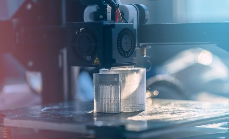
Several printing technologies are used depending on the tissue target. Extrusion-based systems push bio-ink through a nozzle, making them useful for larger constructs such as bone, cartilage, and soft tissue. Inkjet methods deposit fine droplets quickly, which is helpful for thin sheets and cell-patterned surfaces. Laser-assisted techniques offer high resolution and are suited to delicate structures where placement accuracy matters, such as microvessels or nerve-like arrangements.
The field has developed over many years, but recent advances have made the concept more tangible. Researchers have already produced skin grafts, cartilage components, and heart patches that show promise in clinical or preclinical settings. In Spain, bioprinted skin grafts made from a patient’s own cells have been explored for burn care, reducing rejection risk and supporting faster healing. In the United States, printed cartilage has been developed to match knee anatomy and repair damaged joints. In Israel, a bioprinted heart patch containing living cells and vascular structures has been designed to support failing hearts by integrating with native tissue.
These examples matter because they move bioprinting beyond theoretical benefit. They suggest a path toward treatments that are more personalized, less invasive, and better matched to individual anatomy. For burn victims, printed skin can accelerate closure. For joint patients, printed cartilage may restore function. For cardiac patients, living patches could help damaged muscle recover without requiring a full organ replacement.
The medical applications extend well beyond transplantation. Bioprinted tissues can serve as models for disease research, allowing scientists to study cancer progression, immune responses, or tissue injury in a controlled environment. Pharmaceutical companies are also using printed tissues to evaluate drug candidates, which can improve prediction of human response and reduce dependence on animal testing. In personalized medicine, bioprinting could support custom implants, tailored prosthetics, and even patient-specific drug delivery systems that release medication at controlled rates.
The technology is not limited to human healthcare. Veterinary medicine is exploring bioprinted cartilage, skin, and other tissues for animal patients, which can improve recovery and reduce reliance on donor tissue. In the longer term, bioprinting may support long-duration space missions by enabling the production of tissues or replacement parts in environments where supply chains are limited.
Yet the path to routine clinical use is not straightforward. Printing living material requires careful control of cell viability, nutrient supply, waste removal, and mechanical strength. Complex organs need blood vessels, nerves, and coordinated tissue layers, which remain difficult to reproduce reliably. Cost is another barrier, since bio-inks, specialized printers, and expert workflows are still expensive. Ethical questions also need attention, including consent, patient safety, regulation, and the boundaries around creating human-like biological structures.
Despite these challenges, the direction is clear. 3D bioprinting is evolving from an experimental technology into a potential pillar of regenerative medicine. It may not yet print a complete, transplantable heart on demand, but it is already helping to create skin, cartilage, vascular structures, and research models that influence how medicine is practiced. As materials, imaging, and automation improve, bioprinting could transform treatment from replacement with donor tissue to reconstruction with patient-specific living tissue.